What to practice
- reason from molecular structure to biological function
Start with one question
Starch and cellulose are both built only from glucose, yet you digest the starch in a potato and pass the cellulose in its skin untouched. The difference is not the monomer; it is how the monomers are linked and arranged. This chapter covers topics 1.4 to 1.7 of the AP Biology Course and Exam Description. The framework does not ask you to memorize structural formulas. It asks you to reason from structural features, such as branching, saturation, strand direction and side-chain chemistry, to function.
Ask the same questions of every macromolecule
For any biological molecule, ask four questions. What are its building blocks? What covalent bonds join them? Which structural feature matters here? What does that feature let the molecule do? Carbohydrates, nucleic acids and proteins are polymers built by dehydration synthesis (Chapter 4). Lipids are the exception: they are grouped because they are largely nonpolar, not because they share a monomer (Figure 5.1).
| Group | Building blocks | Feature to reason from | Example functions |
|---|---|---|---|
| Carbohydrates | Monosaccharides such as glucose | Linear or branched chains; how monomers are linked | Energy storage; cell walls |
| Lipids | Varied; fats contain glycerol and fatty acids | Nonpolar tails, saturated or unsaturated | Energy storage, membranes, hormones |
| Nucleic acids | Nucleotides | Directional sugar–phosphate backbone; base sequence | Storing and transmitting information |
| Proteins | Amino acids | Sequence and R-group chemistry | Catalysis, transport, structure, signaling |
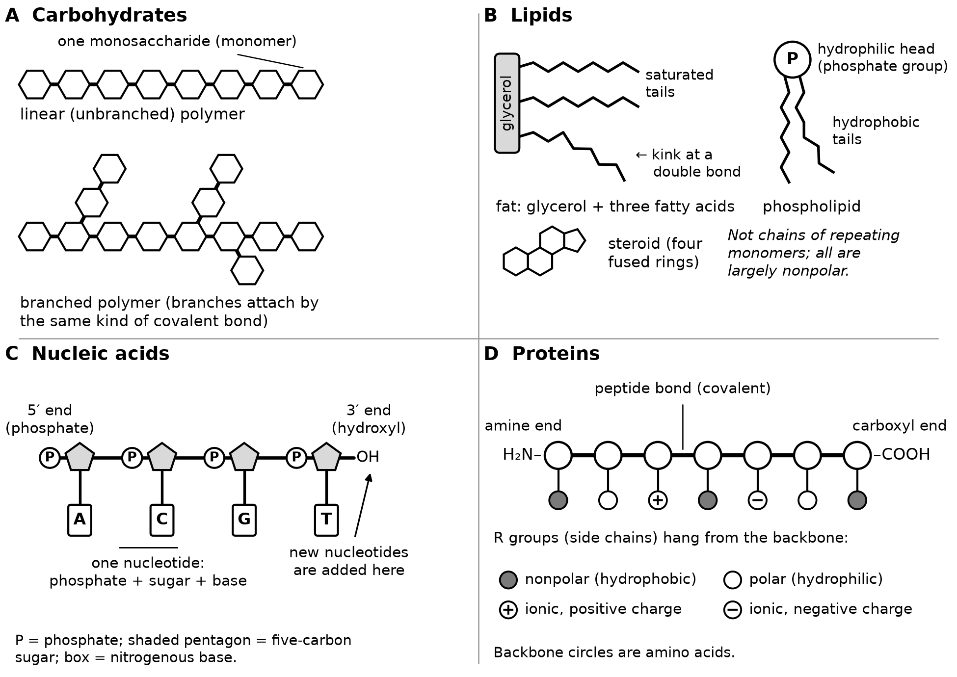
Figure 5.1. How the four groups are assembled. Shapes are symbols, not chemical structures.
Carbohydrates: same monomer, different arrangements
Monosaccharides such as glucose are joined by covalent bonds into polysaccharides, which may be linear or branched. Plants store glucose as starch, a mixture of unbranched chains that coil and branched chains. Animals store glucose as glycogen, which is more highly branched and is kept mainly in liver and muscle. Cellulose, the main component of plant cell walls, is unbranched, and its glucose units are linked in a different orientation. Its straight chains lie side by side and form many hydrogen bonds with one another, building strong fibers. Human digestive enzymes fit the linkages in starch but not those in cellulose (OpenStax Biology 2e, Carbohydrates).
Worked example: predict the rate of glucose release
Prompt. Two storage molecules each contain 2,000 glucose units. Molecule P is one unbranched chain; molecule Q has 80 branches. An enzyme removes glucose only from the tip of a chain or branch. Which releases glucose faster at first?
Reasoning. P has one tip. Q has 81: one for the main chain and one for each branch. Many enzyme molecules can work on Q at once.
Answer. Q. Both store the same amount of glucose; the arrangement controls how fast it can be released.
Lipids: hydrophobic molecules that are not polymers
Lipids consist mostly of nonpolar carbon–carbon and carbon–hydrogen bonds, so they are hydrophobic and do not dissolve well in water (OpenStax Biology 2e, Lipids).
Saturated and unsaturated fatty acids
A fatty acid is a long hydrocarbon tail with a carboxyl group at one end. A saturated tail has no carbon–carbon double bonds, so it carries the maximum number of hydrogen atoms and is straight. An unsaturated tail has one or more double bonds, and each cis double bond bends the tail. Straight tails pack closely and form many attractions with their neighbors, so saturated fats such as butter are solid at room temperature. Bent tails cannot pack closely, so unsaturated fats such as olive oil are liquid (Figure 5.2).
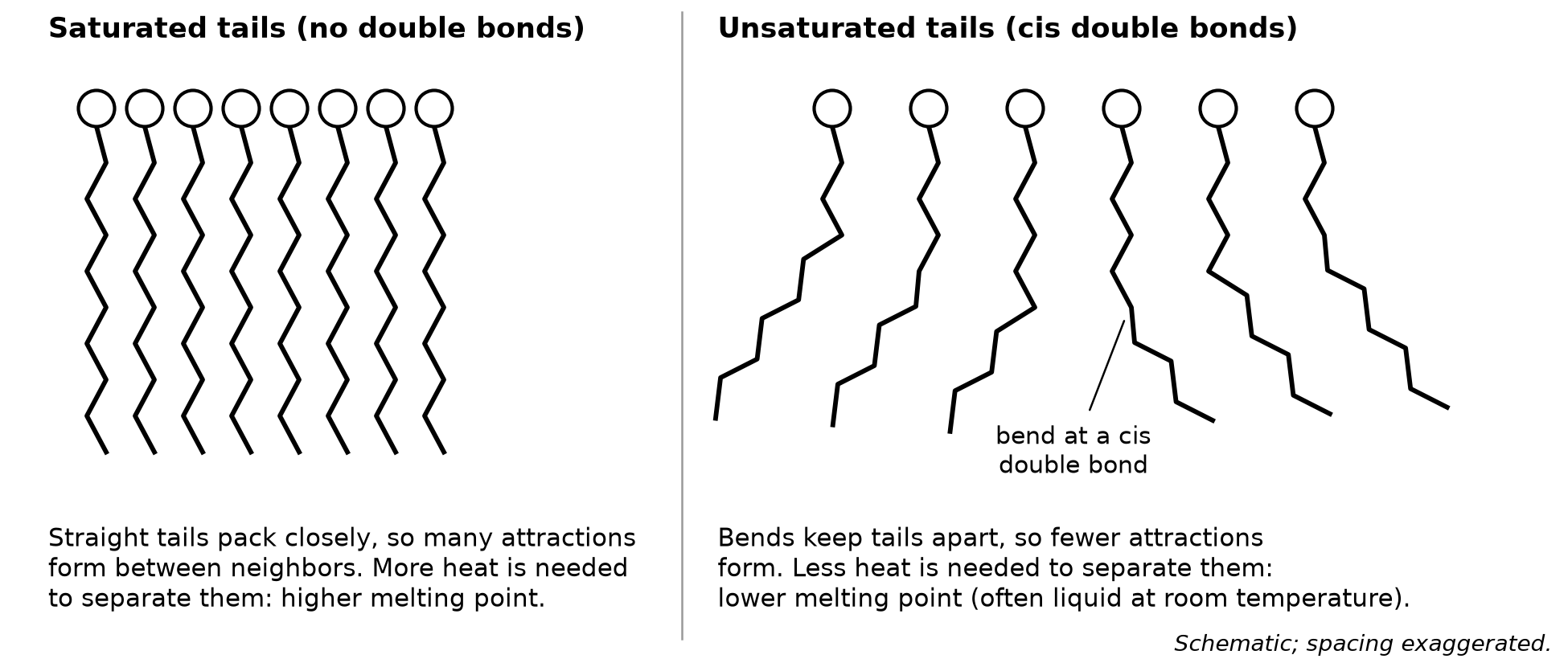
Figure 5.2. Tail shape controls how closely fatty acids pack. Schematic; spacing exaggerated.
The same principle governs membranes: phospholipids with more unsaturated tails make a more fluid membrane. Bacteria grown at lower temperatures build membranes with more unsaturated fatty acids, which keeps membrane fluidity nearly constant across growth temperatures (Sinensky, Proceedings of the National Academy of Sciences, 1974).
Fats, phospholipids and steroids
- A fat is glycerol joined to three fatty acids. Fats store energy for long periods and insulate.
- A phospholipid has glycerol, two fatty acid tails and a phosphate-containing head. The head is hydrophilic and the tails are hydrophobic, so in water phospholipids form a bilayer: heads face the water on both sides and tails face one another inside. Chapter 7 builds membranes from this arrangement.
- A steroid has four fused carbon rings. Cholesterol, a steroid in animal cell membranes, helps regulate membrane fluidity, and steroid hormones such as estrogen and testosterone act as chemical signals.
Worked example: predict melting behavior from tail structure
Prompt. At 24 °C, plant fat F is solid and plant fat G is liquid. Which has more saturated tails? A manufacturer then adds hydrogen atoms across some double bonds in G. Predict the effect on G's melting point.
Reasoning. Solid at room temperature means the tails pack closely, which straight tails allow. Adding hydrogen across double bonds removes bends.
Answer. F has more saturated tails. Hydrogenated G has straighter tails that pack more closely, so its melting point rises.
Nucleic acids: information in a directional sequence
A nucleotide has three parts: a five-carbon sugar, a phosphate group and a nitrogenous base. Covalent bonds join the sugar of one nucleotide to the phosphate of the next, forming a sugar–phosphate backbone with the bases projecting from it. Each strand therefore has two different ends: a 5′ end with a free phosphate and a 3′ end with a free hydroxyl group. New nucleotides are added only at the 3′ end (OpenStax Biology 2e, Nucleic Acids).
| Feature | DNA | RNA |
|---|---|---|
| Sugar | Deoxyribose | Ribose |
| Bases | A, T, C, G | A, U, C, G |
| Usual form | Two strands (double helix) | One strand |
The two strands of DNA run in opposite directions, so they are antiparallel, and their bases pair by hydrogen bonds: A with T and C with G. When RNA pairs with DNA or RNA, A pairs with U. Because every A in double-stranded DNA pairs with a T and every G with a C, the amounts of A and T are equal, and so are the amounts of G and C. The information is the sequence of bases.
Worked example: write a complementary strand
Prompt. Write the DNA strand that pairs with 5′-ATGCCA-3′, labeling both ends.
Reasoning. Pair each base and reverse the end labels, because the partner strand runs the opposite way.
Answer. 3′-TACGGT-5′, which is the same strand written 5′-TGGCAT-3′. An answer of 5′-TACGGT-3′ has the right bases in the wrong direction.
Proteins: sequence determines shape
An amino acid has a central carbon bonded to an amine group, a carboxyl group, a hydrogen atom and a variable R group, or side chain. Some R groups are nonpolar and hydrophobic, some are polar and hydrophilic, and some are ionic, carrying a positive or negative charge. A peptide bond is a covalent bond formed by dehydration synthesis between the carboxyl group of one amino acid and the amine group of the next. A polypeptide therefore has an amine end and a carboxyl end (OpenStax Biology 2e, Proteins).
Four levels of structure
- Primary structure is the sequence of amino acids.
- Secondary structure is local coiling (α-helices) and folding (β-pleated sheets), held by hydrogen bonds between backbone atoms, not R groups.
- Tertiary structure is the overall three-dimensional shape, produced by interactions among R groups. Nonpolar R groups cluster in the interior, away from water; oppositely charged R groups attract; polar R groups form hydrogen bonds; and sulfur-containing R groups form covalent disulfide bridges. Burying nonpolar R groups is a major driving force in folding (Dill, Biochemistry, 1990).
- Quaternary structure is the association of two or more polypeptides, held by the same kinds of interactions. Not every protein has it.
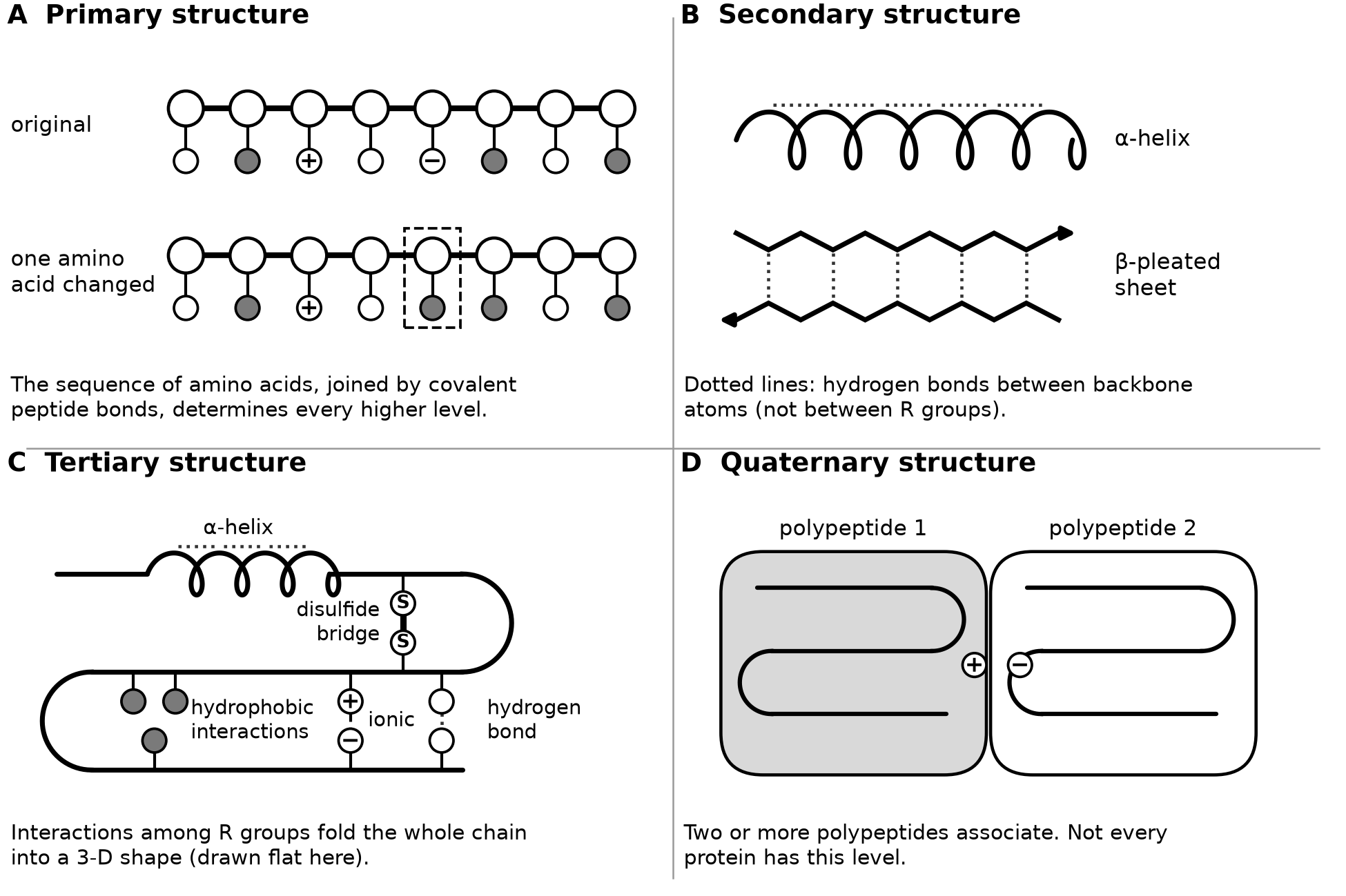
Figure 5.3. The four levels of protein structure. Gray circles are nonpolar R groups, open circles polar ones, and + or − marks a charged R group.
Because shape depends on sequence, one substitution can change function. In sickle cell anemia, valine replaces glutamic acid at one position in each β chain of hemoglobin, a charged R group exchanged for a nonpolar one. The altered hemoglobin molecules form long fibers that distort red blood cells into a sickle shape (OpenStax Biology 2e, Proteins).
Worked example: predict the effect of a substitution
Prompt. On a protein's surface, a positively charged R group attracts a nearby negatively charged one. A mutation replaces the positive R group with a negatively charged R group. Predict the effect, and name data that would test the prediction.
Reasoning. The primary structure changes at one position. Two negative R groups repel, so an attraction becomes a repulsion. The protein's other interactions remain, so structure alone cannot predict the size of the effect.
Answer. The folded shape is likely less stable near that site and could change enough to alter function. Measuring how much heat replicate samples of the normal and mutant proteins tolerate before unfolding would test the prediction.
Common errors to repair
- "Lipids are polymers of fatty acids." Lipids are grouped by being nonpolar; steroids contain no fatty acids.
- "Unsaturated tails have fewer carbons." They have fewer hydrogen atoms because of carbon–carbon double bonds.
- "Starch and cellulose are made of different sugars." Both are glucose; the linkages and arrangement differ.
- "Nucleotides can be added to either end." They are added only at the 3′ end.
- "Secondary structure comes from R groups." It comes from backbone hydrogen bonds.
- "A peptide bond is a hydrogen bond." It is covalent.
Try it
Try it 5.1. Name two structural differences between starch and cellulose and the function each supports.
Try it 5.2. Estrogen, a steroid, crosses a plasma membrane without a channel; glucose does not. Use the bilayer's structure to explain the difference.
Try it 5.3. Write the DNA strand that pairs with 5′-GGATCA-3′, with ends labeled. Then write the RNA strand that would pair with the original strand.
Try it 5.4. A nucleic acid sample is 24 percent A, 16 percent T, 33 percent G and 27 percent C. Is it double-stranded DNA? Explain.
Try it 5.5. Name the level of structure: (a) hydrogen bonds between backbone atoms four amino acids apart; (b) a disulfide bridge between two R groups far apart in one chain; (c) the order of amino acids; (d) four polypeptides that function as one protein.
Check your try-it work
5.1. Cellulose glucose units are linked in a different orientation, giving straight, unbranched chains that hydrogen-bond side by side into strong cell-wall fibers. Starch chains coil or branch, and their linkages fit digestive enzymes, suiting a glucose store.
5.2. Estrogen is largely nonpolar, so it can pass through the hydrophobic tail region. Glucose is polar and is not attracted to that interior, so it needs a transport protein.
5.3. 3′-CCTAGT-5′, also written 5′-TGATCC-3′. The RNA partner is 3′-CCUAGU-5′, also written 5′-UGAUCC-3′.
5.4. No. In double-stranded DNA, A equals T and G equals C, but here A (24) differs from T (16) and G (33) from C (27). The thymine shows it is DNA, so it is probably single-stranded DNA.
5.5. (a) Secondary; (b) tertiary; (c) primary; (d) quaternary.
Review and retrieval
Close the book and rebuild the four-row table from memory, then check it. Tomorrow, redo Try it 5.3 and 5.4 on blank paper. In one week, sketch Figure 5.3 and circle every interaction that involves R groups. These structures return throughout the course: phospholipid bilayers in Chapter 7, enzyme shape in Chapter 8, and DNA, RNA and mutations in Chapters 14 to 16.
Practice
Questions 41–54 and two free-response tasks are printed after this chapter: Written task 2, a nine-point long question built on an experiment, and Written task 3, a four-point short question built on a model. Answer them under timed conditions before checking. Keys, the reasoning behind every choice, and scoring guides with sample responses are under Chapter 5: answers and explanations in the book's answers section.