What to practice
- predict and explain a pathway perturbation
Cells constantly receive chemical messages: a hormone in the blood, a neurotransmitter crossing a synapse, a molecule released by neighboring bacteria. Unit 4 asks you to follow a message from receptor to response and to predict what happens when one step is broken, blocked or stuck "on." You will reuse that prediction skill for gene regulation in Unit 6.
Before you read. Without looking back, write why a large, charged molecule cannot cross a plasma membrane by simple diffusion and what determines a protein's shape. Both ideas return below; check them against Chapters 5 and 7.
How cells communicate: contact, nearby and distant
Topic 4.1 sorts signaling by distance (OpenStax Biology 2e, signaling molecules and receptors).
- Direct contact. Molecules on one cell's surface bind receptors on a touching cell. Immune cells work this way: a T cell inspects antigen fragments displayed on another cell's surface. Adjacent cells can also pass small signaling molecules directly through gap junctions in animals and plasmodesmata in plants.
- Local signaling. A cell secretes a molecule that acts on nearby cells. Growth factors that act on neighbors are local regulators, and neurotransmitters cross the narrow gap of a synapse. Bacteria signal locally too. In quorum sensing, each cell releases a small signaling molecule; as the population grows denser, the molecule's concentration rises until it crosses a threshold and the cells change gene expression together (Miller and Bassler, Annual Review of Microbiology, 2001).
- Long-distance signaling. Hormones travel through the blood in animals or through vascular tissue in plants; ethylene, a gas, can also diffuse between plant parts.
Whatever the distance, a signal changes only cells that have its receptor and the downstream proteins.
Worked example: classify the route, then check the receptor
Beta cells in the pancreas release insulin into the blood. (1) Classify by route: insulin travels in the blood, so it is a long-distance signal. (2) Check reception: a muscle cell without functional insulin receptors bathes in the same insulin but cannot respond. Exposure is not response.
Try it 10.1. Classify each as direct contact, local or long-distance signaling: (a) a neuron releases a neurotransmitter onto a muscle fiber; (b) adrenal glands release epinephrine that alters liver cells; (c) a T cell binds an antigen fragment displayed on an infected cell; (d) a marine bacterium switches on light production only when its population is dense.
Reception: a ligand fits a receptor
The signaling molecule is a ligand. It binds the receptor's ligand-binding domain, whose shape and charge pattern fit only certain molecules; that fit is the source of specificity. Binding changes the receptor's shape, and the change passes to the receptor's intracellular domain, which starts the pathway inside the cell (topic 4.2).
Where the receptor sits depends on the ligand's chemistry:
| Ligand | Crosses the membrane? | Receptor location | Examples |
|---|---|---|---|
| Large or polar, such as peptides | No | Plasma membrane | insulin, epinephrine |
| Small and nonpolar, such as steroids | Yes | Cytoplasm or nucleus | testosterone, cortisol |
Many intracellular receptors become transcription factors once their steroid binds.
Three plasma-membrane receptor types appear often. A G protein–coupled receptor activates a G protein, which is active while bound to GTP and switches itself off by hydrolyzing GTP to GDP. A ligand-gated ion channel opens when its ligand binds, letting specific ions cross, as at many synapses. A receptor kinase is itself an enzyme that phosphorylates proteins when activated (OpenStax Biology 2e, signaling molecules and receptors).
Transduction: relay, amplification and shutoff
Transduction is the sequence of molecular changes that carries the signal from the receptor to the proteins that respond. Three tools do most of the work.
Phosphorylation. A protein kinase transfers a phosphate group from ATP to a target protein, changing its shape and activity. Kinases often activate other kinases in a cascade. Protein phosphatases remove the phosphates and reverse the effect.
Second messengers. Small, nonprotein molecules spread the signal through the cytoplasm. Adenylyl cyclase converts ATP into cyclic AMP (cAMP), and cAMP activates protein kinase A. Phosphodiesterase converts cAMP to AMP, ending that part of the signal. Calcium ions are another common second messenger (OpenStax Biology 2e, propagation of the signal).
Amplification. Each activated enzyme acts on many molecules, so numbers multiply at every enzymatic step. Suppose one receptor activates 20 G proteins, and each activates one adenylyl cyclase that makes 50 cAMP per second. That single receptor yields 20 × 50 = 1,000 cAMP per second. Multiply at enzyme steps; divide when several messenger molecules are needed to activate one protein.
Every pathway also needs an off switch: GTP hydrolysis, phosphodiesterase and phosphatases return it to rest, so the cell can respond to the next signal.
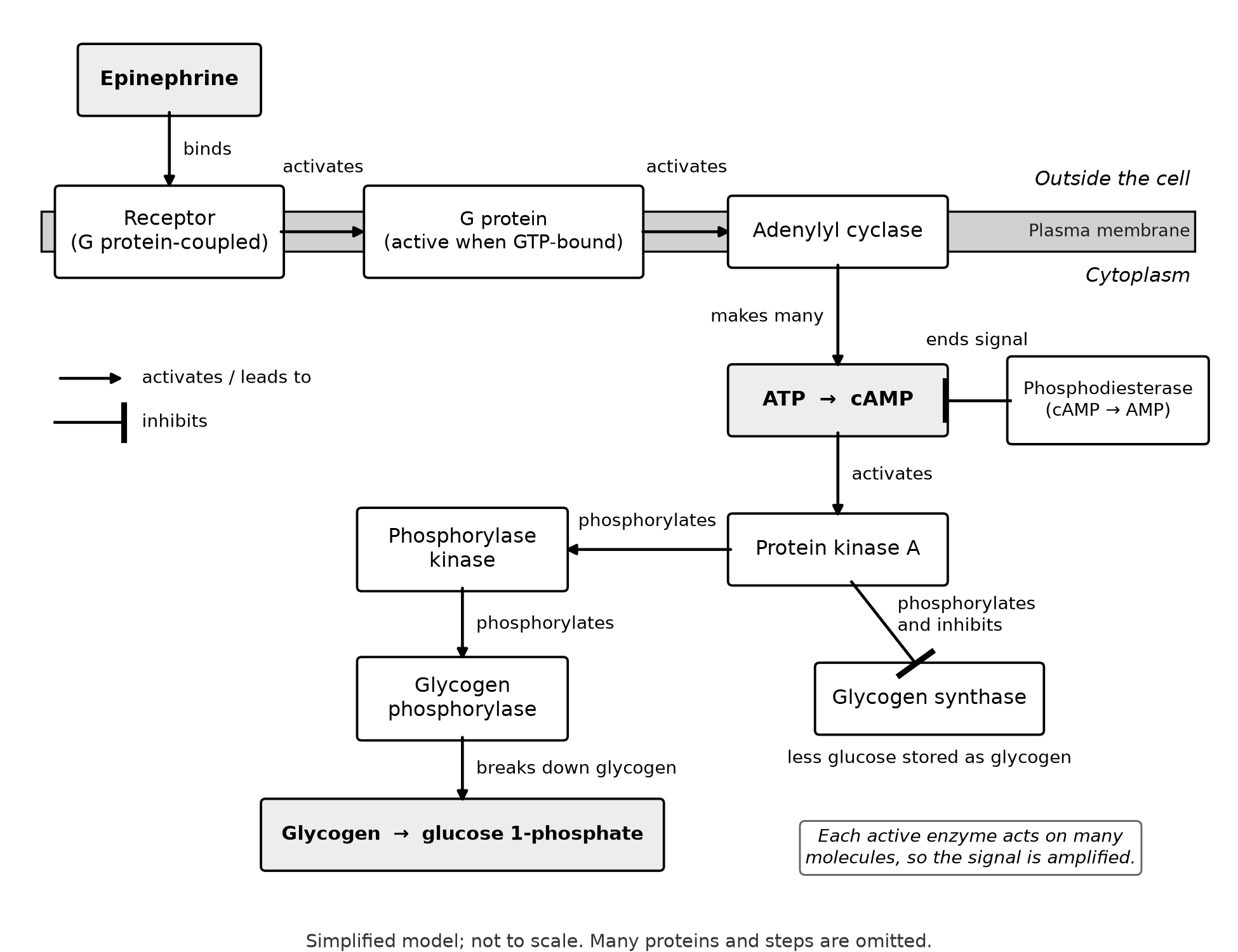
Figure 10.1. A simplified model of epinephrine signaling in a liver cell, drawn from the pathway described in OpenStax Biology 2e, response to the signal. Many proteins and steps are omitted.
Worked example: predict three perturbations
Use one routine for any pathway question:
- Find the changed component in the model.
- Decide what it normally does: activate or inhibit the next step?
- Decide whether the change removes its function or locks it on.
- Trace forward along the arrows, reversing the direction of the effect at each bar-headed link. State the final response, and remember that components upstream of the change are usually unaffected.
A drug blocks phosphodiesterase. Phosphodiesterase normally works against the signal by destroying cAMP. Blocking an inhibitor raises what it inhibits, so after epinephrine binds, cAMP rises higher and lasts longer; protein kinase A stays active longer; glycogen breakdown increases and glycogen synthesis stays inhibited. The receptor and G protein are unaffected.
A mutant G protein cannot hydrolyze GTP. This activator normally switches itself off. Locked in its GTP-bound form, it keeps adenylyl cyclase working after epinephrine leaves the receptor, so cAMP production and glycogen breakdown continue without the signal.
Liver cells from one person release no glucose in response to epinephrine, but a membrane-permeable cAMP analog restores glucose release. The steps from cAMP onward must work, so the defect lies upstream, in the receptor, the G protein or adenylyl cyclase. Had the analog failed as well, the defect would lie at protein kinase A or later.
Common errors.
- Saying that inhibiting an inhibitor lowers the response. Two negatives make a positive.
- Expecting more ligand to rescue a defect downstream of the receptor. Extra signal cannot pass a broken relay.
- Mixing up receptor domains. A changed ligand-binding domain prevents binding; a changed intracellular domain allows binding but blocks transduction.
Responses: what the pathway changes
Topic 4.3 names several kinds of response. Activating existing enzymes, as in Figure 10.1, takes seconds. Changing gene expression takes minutes to hours, because new mRNA and protein must be made. Pathways can also start cell division or trigger apoptosis, programmed cell death that removes unneeded or damaged cells (OpenStax Biology 2e, response to the signal).
Course examples show the range: epinephrine and glycogen breakdown; cytokines that regulate gene expression for cell division; yeast mating pheromones, which bind cell-surface receptors coupled to G proteins (OpenStax Biology 2e, signaling in single-celled organisms); ethylene in fruit ripening; and HOX genes, whose regulated expression lays out the body plan during development.
The same signal can produce different responses in different cells, because those cells carry different receptors or pathway proteins. A mutation in any receptor domain or pathway protein can switch the pathway off or leave it permanently on, and many drugs and toxins work by activating or blocking one specific component.
Try it 10.2. A cytokine binds receptor C, which activates kinase J. Kinase J phosphorylates transcription factor S, and phosphorylated S enters the nucleus and turns on cell-division genes. Protein I binds phosphorylated S and holds it in the cytoplasm. With the cytokine present, predict division-gene expression when (a) protein I is missing; (b) S cannot be phosphorylated; (c) a drug blocks kinase J in cells that lack protein I.
Feedback: hold steady or push to completion
In feedback, the result of a process changes the process itself (topic 4.4).
Negative feedback returns a variable toward its set point. After a meal, blood glucose rises; pancreatic beta cells release insulin; liver, muscle and fat cells take up glucose, and the liver stores glycogen. As glucose falls, insulin release falls. When glucose drops too low, alpha cells release glucagon, which causes the liver to break down glycogen and release glucose (OpenStax Biology 2e, regulation of body processes).
Positive feedback amplifies a change and drives a process to completion. During childbirth, stretching of the cervix triggers oxytocin release, oxytocin strengthens uterine contractions, and stronger contractions increase the stretch (OpenStax Biology 2e, homeostasis). An infant's suckling stimulates oxytocin release, oxytocin ejects milk, and feeding continues (OpenStax Biology 2e, regulation of body processes). In ripening tomatoes, ethylene stimulates the fruit to make still more ethylene (Alexander and Grierson, Journal of Experimental Botany, 2002). Each loop stops when an outside event ends it: birth, the end of feeding, a fully ripe fruit.
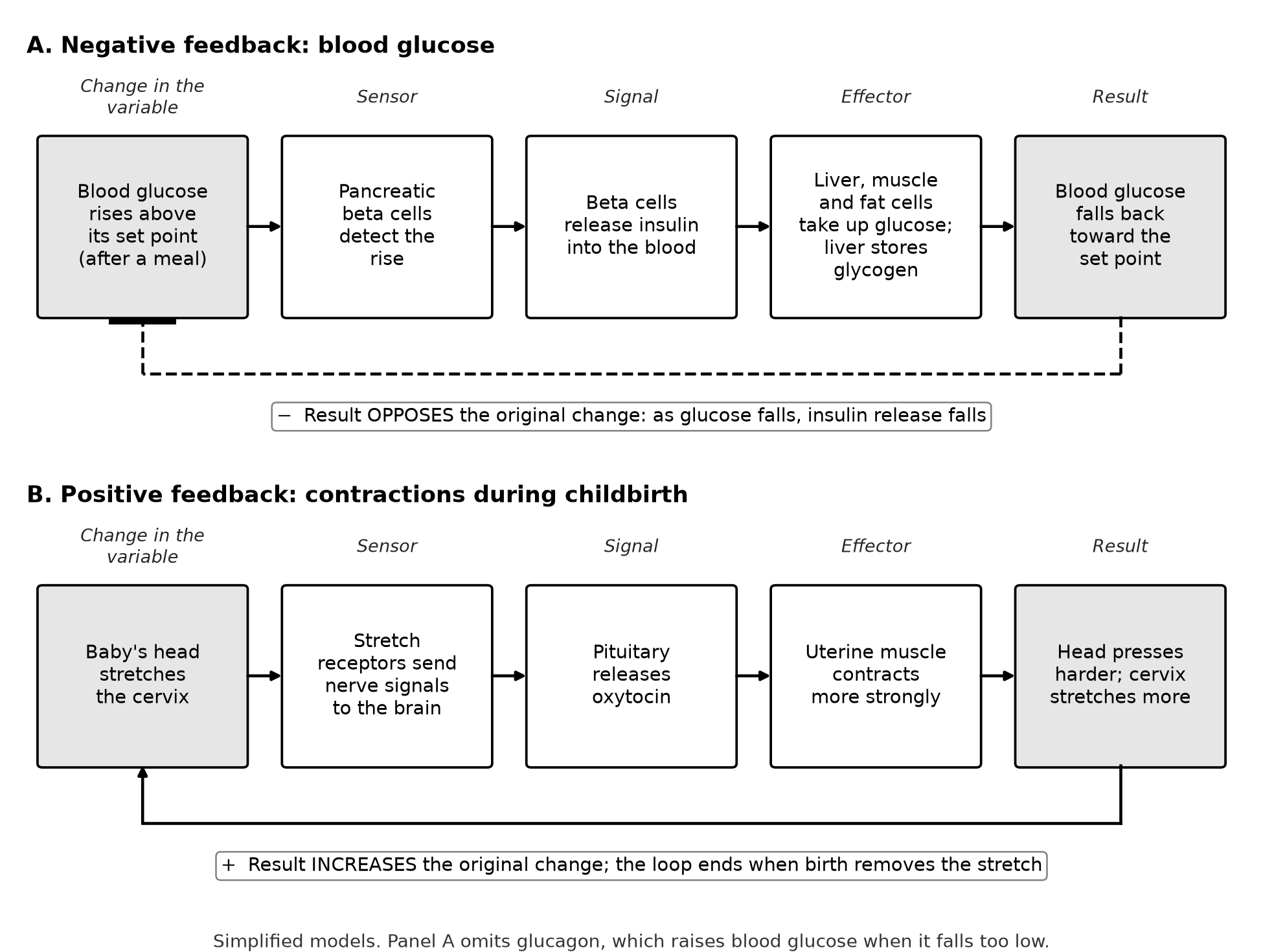
Figure 10.2. The same five parts form a negative feedback loop (A) and a positive feedback loop (B). Panel A omits glucagon, which raises blood glucose when it falls too low.
Two misconceptions cost points. "Negative" does not mean harmful; it means the response opposes the change. "Positive" does not mean healthy or uncontrolled; it means the response pushes in the same direction as the change.
Worked example: predict a failure in a loop
Suppose tissue cells become less sensitive to insulin, one cause of persistently high blood glucose (OpenStax Biology 2e, regulation of body processes). What happens after a meal? The sensor still works: beta cells detect rising glucose and release insulin. The effectors respond weakly, so glucose stays high longer. Because glucose remains above the set point, beta cells keep releasing insulin, and blood insulin may run higher than normal. The loop is still negative feedback: a weak effector changes the size of the correction, not its direction.
Try it 10.3. On a cold day, a person's body temperature falls below its set point. The hypothalamus detects the drop and sends nerve signals that make skeletal muscles shiver, which produces heat (OpenStax Biology 2e, homeostasis). (a) Name the sensor and the effector. (b) Is this negative or positive feedback? Justify. (c) Predict what happens to shivering as temperature returns to the set point.
Check your try-it work
10.1. (a) Local: the neurotransmitter crosses a narrow synapse to a nearby cell. (b) Long-distance: epinephrine travels in the blood. (c) Direct contact: the interacting molecules sit on touching cell surfaces. (d) Local: quorum sensing uses a molecule released among nearby cells.
10.2. (a) Expression rises above normal: nothing holds phosphorylated S in the cytoplasm, so more of it reaches the nucleus. (b) Off: S is never phosphorylated, so it cannot turn on the genes, even with cytokine present. (c) Off: without kinase J, S is not phosphorylated, so removing protein I releases nothing.
10.3. (a) The hypothalamus is the sensor and control center; skeletal muscles are the effector. (b) Negative feedback: heat from shivering opposes the fall in temperature. (c) Shivering decreases and stops as temperature reaches the set point, because the change that triggered it is gone.
Retrieval and review
Today, close the book and redraw Figure 10.1 from ligand to response, marking each link "activates" or "inhibits." Then apply the four-step routine to a drug that blocks protein kinase A. You should predict normal cAMP but little glycogen breakdown, and glycogen synthase no longer inhibited.
In three days, explain without notes why steroid and peptide hormones use receptors in different places, and give one negative and one positive feedback example, naming the event that ends the positive loop. Log any miss in your error log under its topic number, 4.1 to 4.4.
Practice
Chapter 10 practice is Questions 115–130 (multiple choice), Written task 10 (long free response, 9 points) and Written task 11 (short free response, 4 points), printed after this chapter's companion panel with response pages for the written tasks. Answers, reasoning, the error in each wrong choice, scoring guides and sample responses are in Answers, explanations and written responses at the back of the book, under Chapter 10: answers and explanations. After each miss, follow its Repair and retry line.