What to practice
- separate chromosome counts, DNA amount and cell-cycle control
A growth factor can tell a cell to divide, but division is safe only when the cell has copied its whole genome and can split the copies evenly. This chapter covers the cell cycle and mitosis (topic 4.5) and the checkpoints that decide whether a cell moves forward (topic 4.6). Most lost points here come from miscounting chromosomes and from vague answers about checkpoints, so both get worked practice.
Before you read. From Chapter 10, write how a growth factor that never enters a cell could still change which genes the cell expresses. You will connect that pathway to the G1 checkpoint below.
The cell cycle and why mitosis matters
Mitosis transfers a complete genome from a parent cell to two genetically identical daughter cells; it supports growth, tissue repair and asexual reproduction. The cell cycle is the regulated sequence that makes this possible: interphase (G1, S and G2), then mitosis and cytokinesis (OpenStax Biology 2e, the cell cycle).
| Stage | What happens |
|---|---|
| G1 | The cell is metabolically active, grows and duplicates organelles and cytosolic components. |
| S | DNA, still in the form of chromatin, replicates; each chromosome becomes two sister chromatids joined at a centromere. The centrosome, which will organize the mitotic spindle, also duplicates. |
| G2 | The cell replenishes its energy stores and makes the proteins needed to move chromosomes during mitosis. |
| M | Mitosis separates the sister chromatids into two nuclei; cytokinesis then divides the cytoplasm. |
| G0 | The cell leaves the cycle and stops dividing. Many cells can reenter with appropriate cues; mature cardiac muscle and nerve cells generally stay in G0. |
Interphase is not a resting phase. A cell spends most of its time there, and nearly all of its growth and DNA synthesis happen there. Figure 11.1 tracks DNA content through one cycle; read it from left to right and say what happens at each change in the line.
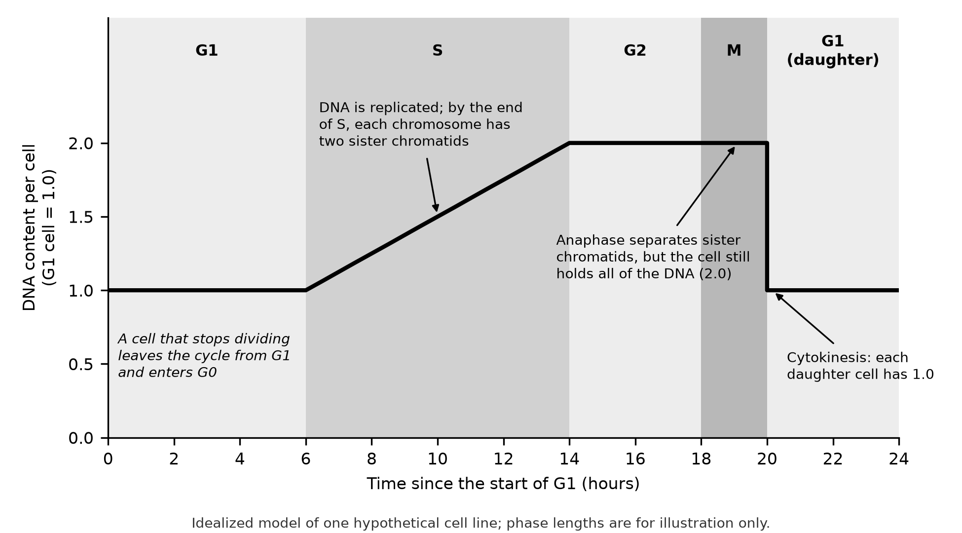
Figure 11.1. DNA content per cell through one idealized cell cycle. Phase lengths are for illustration only; real cells vary widely.
Count four quantities separately
Chromosome questions mix four different quantities. Keep them apart:
- Chromosomes. Count centromeres. Two sister chromatids joined at one centromere are one chromosome.
- DNA molecules. Count chromatids. An unreplicated chromosome has one; a replicated chromosome has two.
- DNA content. The amount of DNA relative to a G1 cell (G1 = 1). It doubles during S and halves only when the cell divides.
- Ploidy. The number of chromosome sets. A diploid (2n) cell has two of each chromosome, one from each parent. Replication does not change ploidy, because sister chromatids are copies, not new sets.
For a cell with 2n = 4:
| Point in the cycle | Chromosomes | DNA molecules | DNA content |
|---|---|---|---|
| G1 | 4 | 4 | 1 |
| G2 or metaphase | 4 | 8 | 2 |
| Anaphase (still one cell) | 8 | 8 | 2 |
| Each daughter cell after cytokinesis | 4 | 4 | 1 |
At anaphase the chromosome number doubles, because sister chromatids separate and each becomes its own chromosome. DNA content does not change: no DNA was made, and the cell has not yet divided. Watch whether a question asks per cell or per nucleus. A cell that has finished telophase but not cytokinesis holds two nuclei, each with DNA content 1, inside one cell with DNA content 2.
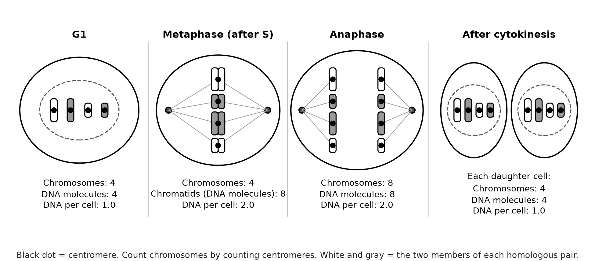
Figure 11.2. Counting through mitosis when 2n = 4. Chromosomes are drawn condensed in every panel so that they can be counted.
Common errors.
- Counting chromatids as chromosomes at metaphase, reporting 8 instead of 4 when 2n = 4.
- Halving DNA content at anaphase instead of at cytokinesis.
- Calling a G2 cell tetraploid. Its DNA content has doubled, but it still has two chromosome sets.
- Pairing homologs at metaphase of mitosis. In mitosis each chromosome lines up independently; homologous pairs form only in meiosis.
Try it 11.1. A plant cell has 2n = 12. Give the number of chromosomes, DNA molecules and relative DNA content (G1 = 1) for (a) G1; (b) metaphase; (c) anaphase; (d) one daughter cell after cytokinesis.
Mitosis step by step
Mitosis alternates with interphase and runs in four steps (OpenStax Biology 2e, the cell cycle):
- Prophase. Sister chromatids condense, the mitotic spindle begins to form and centrosomes move toward opposite poles.
- Metaphase. Spindle fibers align the chromosomes along the cell's equator, each attached to fibers from both poles.
- Anaphase. Sister chromatids separate as spindle fibers pull them toward opposite poles.
- Telophase. The spindle breaks down and a nuclear envelope forms around each set of chromosomes.
Cytokinesis then divides the cytoplasm. A cleavage furrow pinches an animal cell in two; a plant cell, enclosed in a rigid wall, builds a cell plate between the new nuclei instead.
Mitosis and meiosis share the spindle and condensed chromosomes, but mitosis makes two identical cells with the parent's ploidy, while meiosis makes four genetically different cells with half the chromosome number. Chapter 12 develops that comparison.
Worked example: estimate time in mitosis
When cells divide at random times, the fraction of cells found in a stage estimates the fraction of the cycle spent in that stage. A student scores 400 onion root-tip cells and finds 36 in mitosis. The mitotic index is 36 ÷ 400 = 0.09, or 9%. If the whole cycle lasts 20 hours, mitosis takes about 0.09 × 20 = 1.8 hours. The estimate assumes the cells are dividing asynchronously; a treatment that stalls cells in one stage makes that stage look longer than it normally is.
Checkpoints and cyclin–Cdk control
Checkpoints are internal controls that let the cycle continue only when conditions are right. Know the purpose and the timing of each (OpenStax Biology 2e, control of the cell cycle):
| Checkpoint | When | What it checks | Why then |
|---|---|---|---|
| G1 | End of G1, before S | Cell size, energy reserves, growth signals, DNA damage | Passing it commits the cell to dividing, and damaged DNA should not be copied. |
| G2 | End of G2, before M | DNA replicated completely and undamaged | Incomplete or broken copies must not be separated. |
| M (spindle) | Metaphase, before anaphase | Every chromosome attached to fibers from both poles | Separating sister chromatids cannot be undone. |
The idea that a cell confirms one event is complete before starting the next was set out by Hartwell and Weinert, Science, 1989.
Cyclins and Cdks. Cyclin-dependent kinases (Cdks) are present throughout the cycle but are active only when bound to a cyclin. Cyclin concentrations rise and fall during the cycle. When a cyclin accumulates, it binds and activates its Cdk, and the active complex phosphorylates target proteins that move the cell into the next phase. Destroying the cyclin switches the Cdk off. The exam does not require specific cyclin–Cdk pairs or growth factors.
Disruptions. When these controls fail, the result can be cancer or apoptosis. Proto-oncogenes encode positive regulators that promote division; mutated into oncogenes, they can drive division without a signal. Tumor suppressor genes encode negative regulators. One, p53, halts a damaged cell in G1 for repair and can trigger apoptosis if the damage cannot be repaired; p53 is mutated in more than half of human tumors (OpenStax Biology 2e, cancer and the cell cycle).
Worked example: a failed spindle checkpoint
A drug disables the spindle checkpoint in dividing cells with 2n = 4, and one chromosome is attached to only one pole. Predict the outcome. Without the checkpoint, anaphase starts anyway. Both sister chromatids of the poorly attached chromosome can move to the same pole, so one daughter cell receives 5 chromosomes and the other 3. Each daughter now has an abnormal chromosome number and the wrong dose of many genes. A full-credit answer names the checkpoint, its timing (before anaphase), what it detects (attachment to both poles) and the consequence of skipping it.
Try it 11.2. (a) Fibroblasts in culture are moved to a medium with no growth factors. Where in the cycle will most of them stop, and why? (b) Cells with functional p53 are exposed to radiation that breaks DNA. Predict two possible outcomes for a damaged cell. (c) A tumor cell carries a mutated gene whose protein now signals "divide" without any growth factor. Was the normal gene a proto-oncogene or a tumor suppressor? Explain.
Check your try-it work
11.1. (a) G1: 12 chromosomes, 12 DNA molecules, DNA content 1. (b) Metaphase: 12 chromosomes, 24 DNA molecules, 2. (c) Anaphase: 24 chromosomes, 24 DNA molecules, 2. (d) Each daughter cell: 12 chromosomes, 12 DNA molecules, 1.
11.2. (a) Most stop in G1, and many enter G0, because the G1 checkpoint lets a cell proceed only when growth signals are present. (b) The cell arrests in G1, before S, while the damage is repaired; if repair fails, p53 triggers apoptosis. (c) A proto-oncogene. Its normal product promotes division only when signaled; the mutation made an oncogene whose product is always on, a gain of function.
Retrieval and review
Today, draw the cycle as a circle, label G1, S, G2, M and G0, and mark the three checkpoints with what each one checks. Then rebuild the four-quantity table for 2n = 6 without looking.
In three days, explain in two sentences why the M checkpoint must act before anaphase, and why a G2 cell is still diploid. Log any miss in your error log under 4.5 or 4.6.
Practice
Chapter 11 practice is Questions 131–142 (multiple choice) and Written task 12 (short free response, 4 points, with a sketch), printed after this chapter's companion panel with a response page for the written task. Answers, reasoning, the error in each wrong choice, a scoring guide and a sample response are in Answers, explanations and written responses at the back of the book, under Chapter 11: answers and explanations. After each miss, follow its Repair and retry line.