What to practice
- track strand direction and replication evidence
What this chapter trains
This chapter covers topics 6.1 and 6.2 of the AP Biology Course and Exam Description: how DNA and RNA store hereditary information, and how a cell copies its DNA accurately. One habit underlies almost every question here: label the 5′ and 3′ ends before you reason. If the nucleotide structure from Chapter 5 is not yet automatic, reread it first.
Where hereditary information is stored
DNA carries hereditary information from parent to offspring; some viruses use RNA instead. Its organization differs among organisms (OpenStax Biology 2e, DNA structure).
- Prokaryotes typically carry one circular chromosome.
- Eukaryotes carry several linear chromosomes. The DNA is wrapped around histone proteins, which lets a very long molecule be packed and condensed.
- Plasmids are small circular DNA molecules outside the chromosome. They are common in bacteria and also occur in some eukaryotes, such as yeast.
How the double helix stores a copyable message
Each strand has a sugar–phosphate backbone with a 5′ end, which carries a phosphate, and a 3′ end, which carries a hydroxyl group. The two strands run antiparallel.
Bases pair across the helix:
- Adenine pairs with thymine through two hydrogen bonds.
- Guanine pairs with cytosine through three.
A and G are purines, with two rings; C, T and U are pyrimidines, with one. Every pair joins a purine to a pyrimidine, so the helix keeps the same width along its length (OpenStax Biology 2e, DNA structure). Because each base has exactly one partner, either strand specifies the other. That is what makes DNA copyable, and the same pairing rules operate in every known organism.
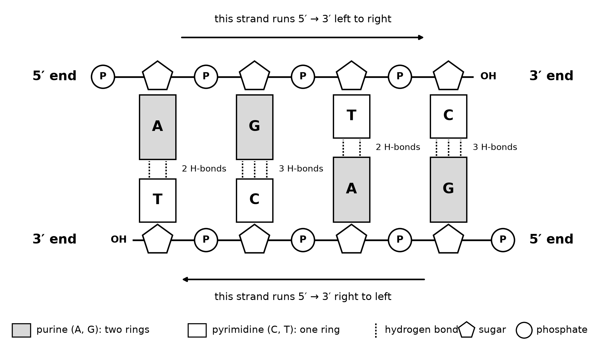
Figure 14.1. Base pairing in antiparallel strands. Original schematic, not to scale.
Worked example: write a partner strand
One strand reads 5′-ATGCCTGA-3′. Write its partner from 5′ to 3′.
- Pair each base in place (A with T, G with C). Directly beneath the given strand, the partner reads TACGGACT. That partner runs 3′ to 5′ from left to right.
- Reverse the order so the 5′ end comes first: 5′-TCAGGCAT-3′.
To check, reverse your answer and pair each base again; you should recover the original strand.
Worked example: base composition
In double-stranded DNA, every A pairs with a T and every G with a C, so A = T and G = C. If a sample is 30% adenine, it is also 30% thymine. That leaves 40% for guanine and cytosine together, so each is 20%. The rule holds only for double-stranded molecules. If A ≠ T or G ≠ C, the molecule is single stranded.
DNA and RNA compared
| Feature | DNA | RNA |
|---|---|---|
| Sugar | deoxyribose | ribose |
| Bases | A, T, G, C | A, U, G, C |
| Usual form | double-stranded helix | single strand that can fold on itself |
| Main roles | stores hereditary information | carries and uses information (mRNA, tRNA, rRNA); genome of some viruses |
Evidence that DNA is the hereditary material
For each classic study below, ask which result rules out the alternative (OpenStax Biology 2e, historical basis).
- Griffith, 1928. Heat-killed disease-causing bacteria, mixed with live harmless bacteria, turned the harmless strain into a disease-causing one. Some substance from the dead cells had transferred a heritable trait.
- Avery, MacLeod and McCarty, 1944. They treated extracts of the dead cells with enzymes that each destroyed one kind of molecule. Destroying DNA abolished transformation; destroying protein or RNA did not (Avery and colleagues, Journal of Experimental Medicine, 1944).
- Hershey and Chase, 1952. They labeled phage protein with radioactive sulfur and phage DNA with radioactive phosphorus. The phosphorus label entered the bacteria and the sulfur label stayed outside, so DNA was the material that entered and directed new phages (Hershey and Chase, Journal of General Physiology, 1952).
Replication is semiconservative
There were three proposed models of DNA copying:
- Semiconservative: each new double helix keeps one parental strand.
- Conservative: the parental helix stays intact.
- Dispersive: old and new pieces are mixed along every strand.
Meselson and Stahl grew E. coli in heavy nitrogen (¹⁵N) and then moved the cells to light nitrogen (¹⁴N). They separated the DNA by density in a centrifuge (Meselson and Stahl, PNAS, 1958).
- After one generation, all the DNA formed a single band of hybrid density. That ruled out the conservative model.
- After two generations, there were equal hybrid and light bands. That ruled out the dispersive model, which predicts one band that keeps getting lighter.
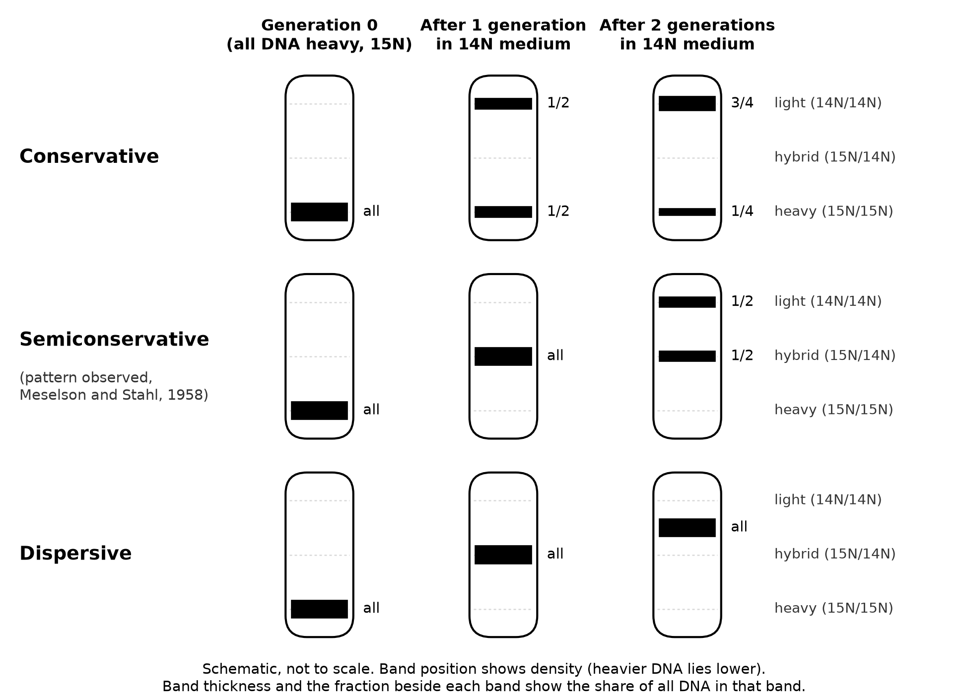
Figure 14.2. Band patterns predicted by three replication models. Original schematic of the experiment's design, not to scale.
Worked example: predict generation 3
Count strands, not bands. The original duplex has two heavy strands, and every later copy keeps one of them. After n generations there are 2ⁿ duplexes, and exactly two of them are hybrid.
After three generations there are 8 duplexes. Two are hybrid, which is 2/8 = 1/4 of the DNA, and the other 6/8 = 3/4 are light. The hybrid share halves each generation but never reaches zero.
Inside the replication fork
Several enzymes work together at the fork (OpenStax Biology 2e, replication in prokaryotes).
- Helicase separates the strands by breaking the hydrogen bonds between paired bases.
- Topoisomerase relieves the over-twisting that builds up ahead of the fork.
- DNA polymerase cannot start a strand on its own. It extends a short RNA primer, adding nucleotides only to a free 3′ end, so every new strand grows 5′ to 3′. Primase, which makes the primers, may appear in diagrams; its name is not required.
- Ligase seals the backbone where neighboring pieces meet, after the RNA primers have been replaced with DNA.
Because the two templates are antiparallel, the two new strands are made differently. The leading strand grows continuously toward the fork. The lagging strand is made in short Okazaki fragments, each growing away from the fork.
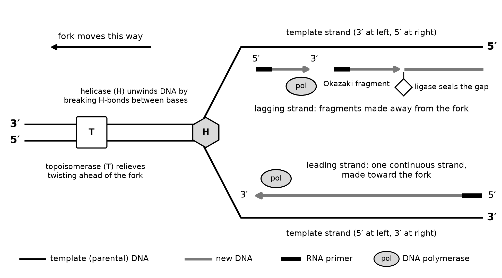
Figure 14.3. One replication fork. Original schematic, not to scale. Primase is not shown.
Worked steps: find the leading strand
Apply these four steps to Figure 14.3.
- Label both ends of each template strand.
- Write the ends of each new strand. It is antiparallel to its template, so a template with 3′ at the left gets a new strand with 5′ at the left.
- A new strand grows toward its own 3′ end.
- If that direction points toward the fork, the strand is leading; if it points away, the strand is lagging.
In Figure 14.3, the upper new strand has 5′ at the left and grows rightward, away from the leftward-moving fork, so it is lagging. The lower new strand grows toward the fork, so it is leading.
Worked perturbation: ligase disabled
Okazaki and colleagues found that DNA made during a brief label was first recovered as short pieces, which were later joined into long strands. This was evidence for discontinuous synthesis (Okazaki and colleagues, PNAS, 1968).
If ligase stops working, new Okazaki fragments are still made, because polymerase and primers are unaffected, but their ends are never sealed. Lagging-strand DNA therefore stays in short pieces instead of becoming long strands.
Origins and speed
E. coli copies its 4.6 million base pairs from one origin in about 42 minutes, with two forks moving apart at roughly 1,000 nucleotides per second (OpenStax Biology 2e, replication in prokaryotes).
Eukaryotic forks move only about 50 to 100 nucleotides per second, but a human genome has up to 100,000 origins, so many bubbles copy it at once (OpenStax Biology 2e, replication in eukaryotes).
Keeping copies accurate
DNA polymerase proofreads: it removes a wrongly paired nucleotide it has just added. Mismatch repair proteins later remove errors that escape proofreading from the new strand. An error that survives both becomes a permanent change in sequence, a mutation, which Chapter 16 develops (OpenStax Biology 2e, DNA repair).
Common errors to repair
- Complementing without reversing. The aligned partner is written 3′ to 5′. Reverse it before you report it 5′ to 3′.
- Saying polymerase "reads" the new strand 5′ to 3′. It reads the template 3′ to 5′ and builds the new strand 5′ to 3′.
- Calling the lagging strand less accurate. It is discontinuous, not careless; its fragments are proofread too.
- Applying A = T to RNA or single-stranded DNA. These equalities come from pairing, so they need two paired strands.
- Calling every enzyme "polymerase." Name the function: unwinding, relieving twist, adding nucleotides or sealing the backbone.
Try it
Try it 14.1. Write the partner of 5′-CGGATTAC-3′ from 5′ to 3′.
Try it 14.2. (a) A double-stranded DNA sample is 34% cytosine. What percent is adenine? (b) A virus genome contains deoxyribose and is 24% A, 31% T, 19% G and 26% C. What can you conclude about its structure?
Try it 14.3. Cells with all-¹⁵N DNA are moved to ¹⁴N medium. (a) Under semiconservative replication, what percent of the DNA is hybrid after four generations? (b) Under the conservative model, what percent is heavy after three generations? (c) After one generation, the DNA forms a single hybrid band. Which alternative to the semiconservative model does that result fail to rule out?
Try it 14.4. A fork moves to the right. The upper template strand has its 3′ end at the left. Which new strand is leading? Where is the newest Okazaki fragment, and which way does it grow?
Try it 14.5. A drug inactivates topoisomerase. Predict the effect on fork movement and explain.
Check your try-it work
14.1. The aligned partner is GCCTAATG, running 3′ to 5′. Reversed, it is 5′-GTAATCCG-3′.
14.2. (a) C = G = 34%, so A + T = 32% and A = 16%. (b) A ≠ T and G ≠ C, so the bases are not paired. The genome is single-stranded DNA.
14.3. (a) 2 of 16 duplexes are hybrid, which is 12.5%. (b) 1 of 8 duplexes is heavy, which is 12.5%. (c) The dispersive model, which also predicts one hybrid band after one generation. A second-generation sample separates the two.
14.4. The upper new strand has 5′ at the left and grows rightward, toward the fork, so it is leading. The lower new strand grows leftward, away from the fork, so it is lagging. Its newest fragment lies nearest the fork, at the right, and grows leftward, back toward the older fragments.
14.5. Twisting builds up ahead of the fork, which makes further unwinding harder, so fork movement slows and eventually stalls. Helicase still works, but the twist it creates is no longer relieved.
Review and retrieval
Close the book and draw a fork with both templates' ends labeled. Mark the leading and lagging strands, a primer, an Okazaki fragment and where ligase acts; check against Figure 14.3. Explain aloud why one hybrid band ruled out only one model. In two days, redraw the fork moving the other way and repeat Try it 14.1 with your own sequence.
Practice
Questions 167–176 and the short Written task 16 follow this chapter's companion panel. They use Figure 14.4, Table 14.1 and Figure 14.5. Answer every question before checking. The keys, explanations, scoring rubric and sample response are in Answers, explanations and written responses, under Chapter 14: answers and explanations. For each miss, write the step from this chapter that would have prevented it.